Hip Dysplasia Basics
CHD is a disorder that begins with joint laxity (looseness)
and then progresses to arthritis over a period of months to years. Its also one of the most common skeletal diseases that vets see. Although the disease is common
in large breeds, it will show up in just about any size/breed. The term basically means "bad development" or "bad growth." Both humans and dogs
get hip dysplasia.
All dogs at birth, have normal hips. However, due to a hereditary disposition and environmental factors, the bones and soft tissues that make up the socket mature
out of sync and the incompatibility displaces normal function in the hip joint. The gene for hip dysplasia is a dominant, co-dominant and also a recessive gene.
(A co-dominant gene is one in which neither gene is dominant). HD is polygenic, meaning it involves many different genes and is influenced by many non-genetic factors.
Therefore you cannot - or should not - breed a dog that carries the gene even though the dog does not manifest dysplasia itself. The dog will either show dysplasia
or carry the genetic information. And this is one of the big reasons why it is extremely difficult to remove the disease from the breed. You get it in your breeding
program when you bred from animals that did not show it.
Dysplasia is not just one disease but many diseases which, when combined, affect the hip joint. It is thought to be a systemic condition, meaning it affects more than
the hips. Because the affected dog shifts its body weight, dysplasia will eventually affect the elbows, shoulders, and the joints between the vertebrae. It is unfortunately,
a common, usually painful and often crippling disease.
The normal hip joint is one of the main weight-bearing joints and is a ball and socket. The ball is on the end of the femur (leg bone) and the socket (acetabulum)
is part of the pelvis. The ball is called the femoral head and fits into the acetabulum in a normal hip. A true ball/socket joint supports movement, has a greater
range of motion than a human's, and has 3 degrees of freedom. The dog's hip joint has a 4th degree of freedom which lets the femoral head move away from the acetabulum.
If the joint has a laxity, the likelihood of dysplasia to develop increases.
The acetabulum is made up of several parts: the ilium (top of the hip), the ischium (lowest part of the hip), the pubis, and the acetabular bone. During the developmental period,
a tight fit is necessary between the acetabulum and the femoral head. The joint should be deep and snug. The first six months of a puppy's life is the most crucial and when the depth
of the socket must be maintained. Early changes aren't easy to detect. Severe cases of CHD can be diagnosed as early as 7 weeks of age. Regardless of what initiated the changes in the
joint, looseness in the joint after 2 weeks of age seems to be a fairly common report preluding CHD. But puppies with tight hips can also develop CHD too.
There is a fibrous structure filled with synovial fluid that surrounds and protects the joint. The cushioning effect of the fluid, the fluid pressure and the elastic nature of the
structure help stabilize the joint as well as keeping out contaminants. Synovial fluid contains the nutrients that are diffused from the blood supply and keep joint tissues alive.
Hyaluronic acid provides lubrication and helps prevents erosion of the cartilage. Dogs with CHD undergo structural changes in the joint, lose hyaluronic acid, and nourishment to the
joint. Then the acetabular rim and the femoral head begin to erode. Tiny micro-stress fractures appear in the bone. The body tries to heal these fissures which makes the acetabulum
fill in and become shallower. The femoral head is pushed further out of the socket, the surrounding supportive tissues weaken and further deform and it becomes a cycle of damage/repair
that leads to a deformed, instable arthritic joint. Perfectly smooth cartilage is gone, lesions develop at the site of abnormal pressure and physical signs appear. The dog has a wobbly gait,
is stiff and sore, gets up slowly, hips do a side/side swinging motion over the hips, knees tuck under the body when sitting, a "click" or "pop" or grating sound as the
dog walks, bunny hopping or a long stride length increases, and/or arched back. Both young and old dogs seem to suffer the most when the weather is cold and damp. The dog then leans forward
to move some of its body weight from the painful hips. The muscles in the thighs atrophy, the hips looks thin and bony, while the shoulder and front leg muscles become overdeveloped. The small
muscles under the scapula overdevelop and the dog's elbows begin to turn outward.
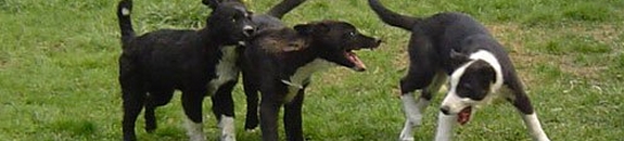
Dogs with CHD have a weak hip joint structure that is more likely to be injured by normal activity like jumping off the couch or out of the car, or playing rough with a playmate.
It may appear as a sudden injury to the owner but the underlying cause is: dysplasia making the joint more susceptible to injury. Puppies should be kept from jumping of engaging in sustained
exercise like jogging, pulling, mushing, running for the first year of their lives.
Influences CHD is diet. A low protein diet and lower activity levels reduced the symptoms of HD. (Protein is thought to be associated with skeletal disease). Dogs predisposed to HD benefit
from a lean diet during their first two years of life. Some vets now feel that slowing the growth rate during the early months of life can lessen the severity of hip dysplasia and possibly
prevent it. Rapid weight gain is associated with HD. Over nutrition increases the risk of HD. Excess calcium is routed to the bones through the influence of calciotropic hormones. Do not supplement
the dog's diet with calcium. Megadoses of ascorbate fed to pregnant bitches and provided to the puppies until young adulthood have been reported to eliminate HD. This is still being studied.
Nutritional management alone cannot manage the development of CHD.
NORMAL HIP Joint: the femoral head fits snugly into the acetabulum.
MODERATE DYSPLASIA: The femoral head flattens, the femoral neck thickens, and the joint becomes loose and unstable.
SEVERE DYSPLASIA: The femoral head and acetabulum are riddled with arthritic and the the femoral head is almost totally dislocated.
Surgical procedures
FHO
Sometimes the head of femur can simply be removed and the surrounding muscles compensate for the missing joint. The dog learns to walk again but running and jumping are not done normally anymore. The best candidates for this type of surgery are smaller dogs. Large and heavy dogs will still have pain. The goal of this type of surgery is to eliminate pain by removing the point of contact in the joint. a false joint is made from the scar tissue that develops. This procedures usually works for dogs under 50 pounds. How successful the FHO is depends on technical skill, the weight and age of the dog, amount of atrophy before surgery, and therapy after surgery.
Sometimes a muscle flap is used to create the scar tissue and support or cushion the femoral shaft. The muscle sling is used when the dog weighs more than 50 pounds and is usually either the biceps femoris muscle or the gluteal muscle. When the muscle used is the rectus femoris muscle, as a sling, the dog is more obviously lame, return to function is slower and there is no improvement in the range of coxofemoral motion.
The thigh muscle shows more atrophy. The muscle is sewn in
place and and secured to the acetabulum and used to pad the area between the shaft and the pelvis. Gradually that piece of muscle dies as a muscle and becomes a fibrous mass that absorbs impact. When the a partial thickness of the biceps femoris muscle is used as a sling, the dog should have more improvement in limb function and range of motion than if a deep gluteal muscle were used as a muscle sling.
After the femoral head has been cut off, the surgeon checks the range of motion and makes sure there are no obstructions to normal movement. If there is a dry crackly sound, not enough of the femoral neck was removed, or there could be bone fragments still in the site. Either of these will cause pain and loss of function. The removal of the femoral head shortens the leg slightly and causes the leg to draw inward.
The FHO is strictly considered and end-salvage procedure. Only dogs with severe bony changes should be considered for this procedure. Bed sore type of ulcers are frequent problems associated with this type of procedure. Best results are with dogs under 35 pounds. Although this doesn't return the hip to normal, it should relieve the pain of arthritis. Its simple, has few complications and can be done at any age. On rare occasions the ischiatic nerve gets entrapped in the muscle that is used for the sling. The dog has pain and a portion of the ischium has to be resected in order to provide relief.
TPO
This surgery, to be effective, has to be done before arthritis deforms the acetabular rim or the femoral head. The best candidate is a young dog between 5 months and 1 year of age. In this procedure the acetabulum is rotated to a more normal position in relation to the femoral head, and puts it deeper into the socket by breaking it into three pieces. The new position is stabilized with a plate and bone screws. the hips is able to develop more normally without developing arthritis. About 90% of dogs get good to excellent results and live a normal life of activity. If a TPO is done on the second leg, it is usually done 3-6 weeks later.
THR
THR is currently the best available treatment for severe hip dysplasia in large dogs. Success rate is over 95% and dogs with successful implants are able to preform almost any task done by dogs with normal hips. Therefore, it is the treatment of choice for dogs used for working or sporting activities or when optimal hip function is desired. Most owners report the dog's personality improves because the pain is gone. Most dogs resume normal level of activity within 2 months after surgery.
Total Hip replacement involves removing the ball and the socket portions of the hip joint and replacing them with artificial ones. The femoral head is cut off. The inside of the femur is drilled out and reamed with a tapered reamer, then finish filed and broached. The femoral stem is test-fitted and then cemented in place. Often the glue includes and antibiotic. The acetabulum is reamed, the acetabular cup is fitted and cemented into place. The hip components are in three units allowing for mix and match to better match the dog's original skeletal conformation.
Possible complications exist. Infection is the most common occurrence. Madison vet hospital boasts a 1% rate. Second possible complication is called "cement disease" or aseptic loosening. a Fibrous membrane normally develops between the bone and the cement. With cement disease, a synovial like membrane containing particulate debris develops that produces large amounts of bone resorbing factors. Another complication could be sciatic neuropraxia (inability of the nerve to conduct impulses).
The dog must be fully grown (over 9 months old with fully developed growth plates), over 40 pounds, free of infections or neurologic disorders. They must be healthy. Strict confinement and close supervision is required for 8 weeks after surgery. 80% of dogs only need one side repaired. Ultimately most dogs regain full, pain free function after a THR. The most painful hip is replaced first and if the second hips needs to be replaced, it is at least 2 months later. Most dogs walk on their new hip immediately after surgery. Recovery time is quicker if the THR is done before the hind leg muscles have atrophied. |